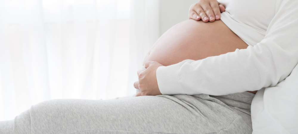
When an unborn baby has an abnormality, decisions about the delivery and neonatal care can often be planned in advance – with the help of an MRI.
However, MRI image acquisition can be tricky and quality reporting is an acquired skill. Dr Lauren Raubenheimer is an expert in the field, having honed her skills in London under word-renowned fetal and neonatal imaging specialist, Prof. Mary Rutherford. She recently joined SCP Radiology as a consulting radiologist and provides insights into fetal MRI and what it can mean to expecting couples and their healthcare providers.
‘When a couple discovers the baby that they carry has an abnormality, the impact is enormous and can be life changing. As a mother, I have a huge emotional investment in my work. I hope that my findings give parents more answers as they navigate a difficult path.’
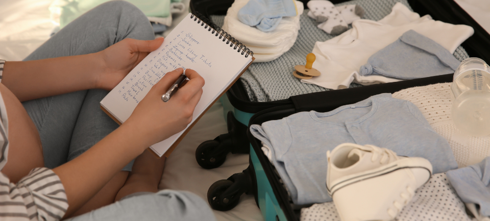
She says that, in many cases, the timing and route of delivery, as well as the neonatal care that will be required, can be planned in advance with help from an MRI. ‘Nothing is better than confirming a minor abnormality, with a good prognosis and giving parents some reassurance.’
Here she talks about the how, why and when a fetal MRI is indicated and the safety factors taken into consideration when doing so.
When would fetal MRI be recommended?
Patients are typically referred from Fetal Medicine Specialists after an abnormality has been detected on a screening ultrasound. An MRI can be done to confirm the abnormality, as well as to assess for abnormalities that are not readily visible through standard imaging techniques. This can significantly affect the prognosis.
A fetal MRI can be performed either in the second or third trimester. ‘My special interest is in developmental fetal brain abnormalities but I also perform MRIs for body abnormalities, including congenital diaphragmatic hernia, congenital lung lesions, spina bifida, kidney anomalies and fetal tumours.’
Why not ultrasound?
MRI can be superior to an ultrasound in certain instances. Such as assessing the folding of the brain and assessing parts of the brain obscured by the skull, when amniotic fluid is low (amniotic fluid is a clear to slightly yellow liquid that cushions a fetus within the amniotic sac) and when mothers have a high body mass index (BMI).
How are quality images obtained?
Fetal movement has previously been an obstacle in getting good quality MR images in the past. ‘But with today’s magnets and the use of faster imaging sequences, we can obtain excellent image quality, she explains.
That said, a very busy baby calls for considerable skill and patience from the radiographer and, at SCP Radiology, fetal MRI scans are done exclusively by the lead MRI radiographer, Andrea Nagel.
Safety comes first
Safety is of utmost importance. Present data shows ‘no conclusively documented harmful effects of MRI imaging on the developing fetus, providing it is at the safe and optimal level (1.5 T”)’.
For the peace of mind of expectant parents, Dr Raubenheimer adds that MRI does not use ionising radiation and intravenous contrast is not administered in fetal MRI. ‘By working within strict parameters, potential harm to the fetus is prevented’.
About referral and funding
Fetal MRI is covered by medical aids but, as is the case with all other MRI scans, preauthorisation is required..
It is preferable that patients are referred from a Fetal Medicine Specialist after a detailed ultrasound. ‘Having access to ultrasound reports and knowing the exact gestation is crucial to an accurate MRI report’, Dr Raubenheimer emphasises. In general, she is in close communication with her colleagues in fetal medicine when it comes to individual patients.
Parents who want to read more about the safety of and preparation for fetal MRI, can download SCP’s information sheet from the website: www.scp.co.za
About Dr Lauren Raubenheimer
Dr Raubenheimer obtained her MBChB with first class honours from the University of Cape Town and her radiology qualification with distinction from the same institution.
After graduating, she worked in both the public and private sectors in South Africa and developed a special interest in fetal imaging and neuroradiology. She enrolled for the European Diploma in Neuroradiology (EDiNR) and, in 2019, did an observership in fetal and neonatal brain MRI at the St Thomas’ Hospital Centre for the Developing Brain in London, under the guidance of Prof. Rutherford.
Since then, she has reported fetal MRIs for Groote Schuur Hospital, as a volunteer medical specialist from 2019 to 2023 as well as private-sector fetal MRIs, first in Stellenbosch and more recently at Mediclinic Cape Gate in partnership with SCP.
Aside from her fetal imaging work, she also currently does contract reporting for an international radiology provider.
targeted toward Parents.
We understand that there are many aspects that encompass a Mother, Father or Child and strive toward providing resources and services that accommodates
this.
Our content is aimed to inform and educate families on issues starting from
pregnancy through to the challenges of the teen-age years.
- How to Talk to Your Kids About Tough Topics - October 15, 2025
- Creative Ways to Sneak Veggies into Kids’ Meals - October 14, 2025
- Turning Chores into Fun Games for the Whole Family - October 13, 2025