
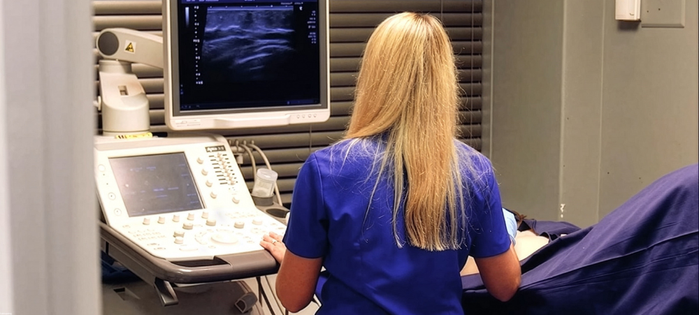
One of the most important and least understood factors in breast cancer detection, is breast density. Dr Marna du Toit, a radiologist from SCP Radiology who recently obtained a European Diploma in Breast Imaging (EDBI), explains what dense breasts are, the impact on the kind of imaging that will be used and explanations of each one.
Breast cancer remains the most commonly diagnosed cancer in women globally and multiple factors determine a women’s risk of being diagnosed. One of which is dense breasts. Dr du Toit says, ‘It is not only an independent risk factor but also has an impact on breast cancer screening.’
Breast tissue density is plotted on a typical Bell curve:
- Type A – 10%: Almost entirely fatty
- Type B – 40%: Fatty with scattered areas of density
- Type C – 40%: Dense with scattered areas of fatty tissue
- Type D – 10%: Extremely dense
What do dense breasts actually mean?
Your breasts are composed of fibrous tissue (supports and holds tissue in place), glandular tissue (milk glands which produce milk and ducts that transport the milk) and fatty tissue (fills the space between fibrous and glandular tissue). If you are told that you have dense breasts – referred to as ‘fibroglandular density’ – it means that you have more fibrous and glandular tissue and less fatty tissue.
How many women have dense breasts and why?
Having dense breasts isn’t something you can control, it is often inherited but can change over time. Some women are more likely to have dense breasts, such as those who are pre-menopausal, are menopausal and taking hormone replacements or are lean with low Body Mass Index (BMI).
How does this impact breast screening?
Dense breast tissue can make breast cancer more difficult to see or detect on a mammogram, for the following reasons:
- Breast cancer can show up in many different ways but often appears white. On a mammogram, with mostly dense tissue which shows as white, it may be harder to detect which increases the risk that cancer may be missed. It’s far easier if it’s surrounded by darker fat (which is reflected as the dark/black areas on a mammogram). High breast density reduces the imaging sensitivity from 86-89% in fatty breasts (Type A) to 62-68% in extremely dense breast tissue (Type D)
- There is a 2-3% higher risk of developing breast cancer
Average woman lifetime risk of breast cancer: About 2-13% . Women with Type D breasts: Often estimated about 18-25% lifetime risk.
- However, breast density alone does not predict the risk of breast cancer, it is just one piece of the puzzle
‘This means you probably need supplemental imaging in addition to a normal digital mammogram,’ explains Dr du Toit.
Digital mammogram
A digital mammogram is the primary imaging method for screening. It uses low-dose X-rays to take detailed pictures of the breast tissue, to check for the possible presence of breast cancer and benign breast disease.
Why it’s useful
A mammogram can pick up breast cancer before any symptoms develop and detect a tumour that cannot be felt – this means less aggressive treatment and a reduction in mortality rates.
Something to consider: Younger women often have more dense breast tissue, which means changes in the breast might be obscured. Therefore, a mammogram is not used as a screening tool in women under 40 but can be used diagnostically.
Dr du Toit says there is a difference between a screening mammogram, which looks for breast cancer before there are any symptoms or a diagnostic mammogram which investigates a specific symptom or abnormality.
When to start
Average risk: Annual screening starting at age 40 and continuing for as long as you are in good health. If you have a first degree relative diagnosed at a young age, speak to your doctor about starting your screening earlier.
‘There are various supplemental screening methods used for women with dense breasts. These include: Ultrasound, a 3D mammogram (tomogram) and an MRI’, explains Dr du Toit.
A 3D mammogram (tomogram)
This captures multiple X-ray images, from different angles, to create a series of very thin cross-sectional ‘slices’ of the breast. A radiologist will scroll through these images to look for areas of distortion, which is often an early sign of cancer before other symptoms appear.
Advantages: It is helpful for women with dense breasts because it is a clearer picture than a 2D mammogram and improves detection rate.
Something to consider: It does have a slightly higher radiation dose than a regular mammogram but it is still a safe dose ascribing to the ALARA (As Low As Reasonably Possible) rule in radiology.
Ultrasound
Ultrasound is a non-invasive procedure that uses sound waves to create images – particularly to investigate lumps felt by hand or seen on the mammogram.
Advantages: It is especially helpful in younger women with dense breast tissue, as it can help detect cancers that may not be visible on standard mammograms. It provides a real-time view of the breast during the scan and is excellent for examining the lymph nodes in the underarm area. There is no radiation involved.
Something to consider: It may not detect all cancers, particularly small lumps or micro-calcifications, which can be early signs of cancer.
Magnetic Resonance Imaging (MRI)
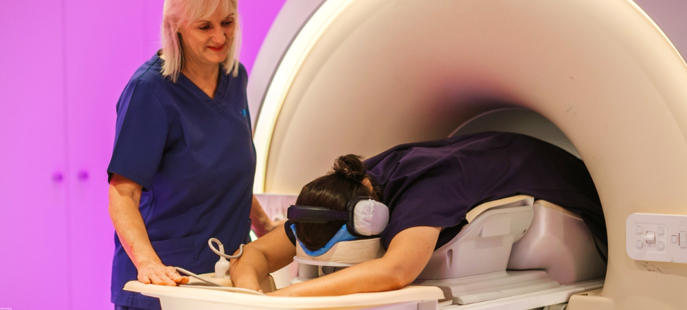
A breast MRI uses magnetic fields and radio waves to create detailed pictures of the breast. It can be used together with or instead of a mammogram. You do, however, need an injection of contrast dye. This is the ‘Gold Standard’ in breast imaging and has a 90% sensitivity. Can be used as screening in young or high-risk patients, diagnostic imaging, pre-operative to determine the extent of disease and breast cancer surveillance.
Advantages: No radiation is used. It’s a good screening tool for high-risk individuals and patients with extremely dense breasts, as well as those with a strong family history and gene mutations.
Something to consider: You may have an allergic reaction to the contrast dye containing gadolinium. This is very rare, usually mild and quickly treated. Most reactions are mild (nausea, warmth, flushing, dizziness, mild urticaria/itching) and don’t last long.
Dr du toit says that knowing your breast density and the implications is crucial for assessing your overall breast cancer risk (and should be included on your imaging report). It also determines if you could benefit from supplemental screening tests – such as a breast ultrasound or MRI – to ensure early detection. The European Society of breast screening advises women aged 50 to 70 years, with extremely dense breasts, to have a breast MRI every 2 to 4 years.
‘Remember these tests can detect breast cancer long before you can feel a lump. Early detection is crucial,’ says Dr du Toit. ‘It can save your life.’

We understand that there are many aspects that encompass a Mother, Father or Child and strive toward providing resources and services that accommodates this.
Our content is aimed to inform and educate families on issues starting from pregnancy through to the challenges of the teen-age years.
- You Look After Everyone Else. Isn’t It Time You Put Yourself Back on the List? - September 17, 2026
- How breast density matters in breast cancer - September 17, 2026
- 222 Nights of Hope: Body20 Eases the Journey for Families Facing Childhood Cancer - September 16, 2026