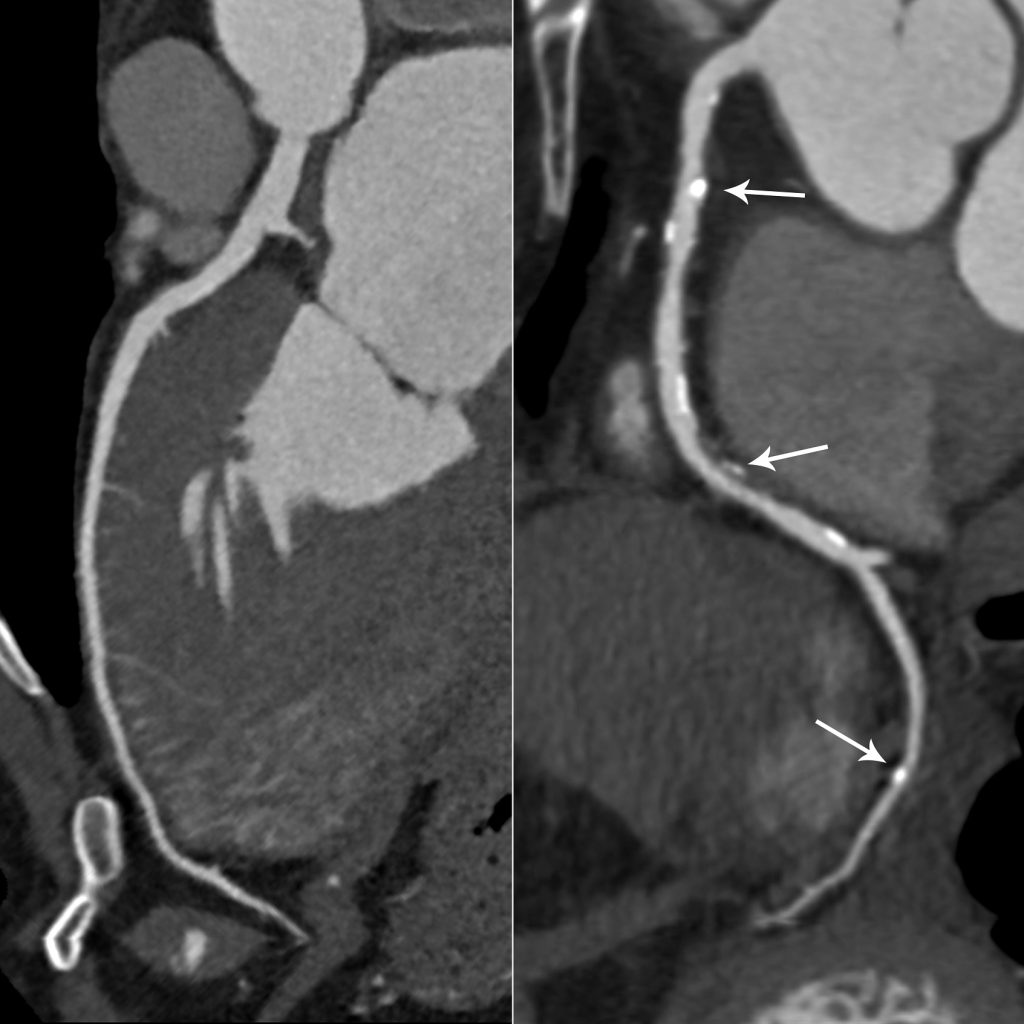
Radiology imaging techniques play a crucial role in the early diagnosis, management and monitoring of lifestyle diseases. Advancements in high-quality imaging, using reduced radiation doses, have positioned radiology ideally for this role. This contributes significantly to the understanding and treatment of these conditions.
One such condition is cardiovascular disease, which accounts for approximately 30% of deaths worldwide, coronary artery disease is the commonest form. Dr Vishesh Sood, a diagnostic radiologist practising at SCP Radiology, says ‘radiology’s role in diagnosing and managing coronary artery disease is pivotal.’
What is coronary artery disease?
Coronary artery disease is the gradual narrowing of the coronary arteries, which supply blood to your heart.
What causes this narrowing?
It is caused by plaque building up in the inner lining – a process known as atherosclerosis. Plaque is made up of deposits of varying degrees of fatty substances, cholesterol, cellular waste products, calcium and fibrin. As it builds up in the arteries, the artery walls become progressively thickened and stiff.
What happens when this build up occurs?
These signs and symptoms of coronary artery disease occur when the heart doesn’t get enough oxygen-rich blood. Reduced blood flow to the heart can cause a wide range of symptoms, most commonly chest pain (angina) and shortness of breath. A complete blockage of blood flow can cause a heart attack, which means that blood flow to a part of the heart is reduced for a long enough time and the heart muscle is permanently damaged or dies.
What are the risk factors you CAN control?
- High blood pressure
- Elevated cholesterol levels
- Smoking
- Diabetes
- Overweight or obesity
- Lack of physical activity
- Unhealthy diet and stress
What you can’t control
However, it’s important to note that genetics also play a crucial role in determining an individual’s risk level. Managing coronary artery disease involves adopting lifestyle changes such as a healthy diet, regular exercise, and quitting smoking, along with medications like statins to lower cholesterol. A coronary CT empowers doctors to recommend these changes and treatments, with the aim of preventing serious cardiovascular events
How long does this condition take to develop?
Coronary artery disease generally progresses gradually over decades. The disease may go entirely unnoticed, until a significant blockage causes problems or a heart attack occurs.
‘Which is why,’ says Dr Vishesh Sood, ‘we advocate the use of a CT Coronary Angiography, the most sensitive imaging method to confirm the disease.’
He says, ‘This is particularly important in patients with non-specific symptoms, for example chest pain, which could be caused by a range of problems like musculoskeletal issues, gastrointestinal conditions or even anxiety. In these instances, coronary CT angiography is an excellent non-invasive test to confirm whether high levels of plaque are present in the coronary arteries.’
What is a CT Coronary Angiography (CTCA)?
It produces detailed images of the heart for precise evaluation of the coronary arteries. A contrast dye is injected into a vein (usually in the arm) before imaging, to increase the visibility of any obstructions in the coronary arteries.
 The advantages
It provides high resolution images of the coronary arteries, during a short, non-invasive procedure.
The disadvantages
Even though CTCA provides detailed images of the coronary arteries, it does not allow for interventions, such as inserting a stent during the same procedure. It is used primarily for diagnosis.
Dr Sood says, ‘This is not the only method for detecting coronary artery disease. The diagnostic ‘gold standard’ has always been coronary/catheter angiography.’
What is a Coronary (Catheter) Angiography
During this procedure, a catheter is threaded through blood vessels to the coronary arteries and a contrast dye is injected to make the arteries visible on X-ray images.
The advantages
It allows for detailed analysis of the blood flow within the coronary arteries. If a balloon needs to be inserted to stretch open a narrowed or blocked artery, it can be done at the same time. ‘Although’, says Dr Sood, ‘the more popular choice is to insert a permanent stent to allow blood to flow more freely, which can also be performed during a Coronary Angiography.’
The disadvantages
It is an invasive procedure. Threading a catheter through blood vessels, usually from the groin or wrist, up to the coronary arteries can be uncomfortable for patients. It may take an hour or longer to complete and may result in bleeding, infection and some potential damage to blood vessels.
Who makes the decision to do either procedure?
The specialist will do a thorough assessment and then makes the decision. ‘The choice between cardiac CT and angiogram depends on the specific situation, the information needed and individual patient factors,’ explains Dr Sood. ‘Cardiac CT is often used for initial screening non-invasive assessment, while conventional coronary angiography is usually for cases where more detailed information about the blood flow is required or when the plan is to insert a balloon or stent into the artery to expand it.’
CTCA is quickly becoming a cornerstone in modern cardiology to detect coronary disease can be detected and managed before it becomes life threatening.
We understand that there are many aspects that encompass a Mother, Father or Child and strive toward providing resources and services that accommodates this.
Our content is aimed to inform and educate families on issues starting from pregnancy through to the challenges of the teen-age years.
- Say Hello to the Ultimate Holiday Brunch Bite - December 17, 2025
- Tiny Toons Looniversity Returns: Meet the Voice Behind Plucky and Hamton! - December 12, 2025
- From Pain to Possibility: Panado®’s New Marketing Campaign, Highlights The Joy Of Pain Relief - December 10, 2025


 The advantages


